Lysine | C6H14N2O2 | Amino Acid Database

Generic NameIDType
LysineAADB0012Essential
Function: The essential amino acid l-lysine (Lys) is needed for the synthesis of peptides and proteins and as a precursor of carnitine. Use as an energy fuel requires adequate supplies of thiamin, riboflavin, niacin, vitamin B6, pantothenate, lipoate, ubiquinone, iron, and magnesium. Food sources: Adequate amounts are consumed when total protein intake meets recommendations. Dietary supplements containing crystalline Lys are commercially available. Requirements: Estimates of daily Lys requirements vary, but they appear to be near 30mg/kg in healthy adults (EI-Khoury et al., 2000). Deficiency: Prolonged lack of Lys, as with all essential amino acids or a lack of protein, causes growth failure, loss of muscle mass, and organ damage. Excessive intake: A very high intake of protein and mixed amino acids (i.e., more than three times the RDA, which is 2.4g/kg) is thought to increase the risk of renal glomerular sclerosis and accelerate osteoporosis. There have been anecdotal reports of symptoms alike to EMS and other severe illnesses following the use of some synthetic Lys preparations (Kohlmeier 2015)
Amino acid ClassSide Chain Type
BasicPolar, positively charged
Kohlmeier, M. (2015). Nutrient metabolism: Structures, functions, and genes (2nd Edition). Academic Press. 10.1016/C2010-0-64980-7
DrugBank
Lysine (abbreviated as Lys or K) is an α-amino acid with the chemical formula HO2CCH(NH2)(CH2)4NH2. This amino acid is an essential amino acid, which means that humans cannot synthesize it. Its codons are AAA and AAG. Lysine is a base, as are arginine and histidine. The ε-amino group acts as a site for hydrogen binding and a general base in catalysis. Common posttranslational modifications include methylation of the ε-amino group, giving methyl-, dimethyl-, and trimethyllysine. The latter occurs in calmodulin. Other posttranslational modifications include acetylation. Collagen contains hydroxylysine which is derived from lysine by lysyl hydroxylase. O-Glycosylation of lysine residues in the endoplasmic reticulum or Golgi apparatus is used to mark certain proteins for secretion from the cell.
Descriptions
ChEBI
ChEBI :16977

L-lysine is an L-alpha-amino acid; the L-isomer of lysine. It has a role as a micronutrient, a nutraceutical, an anticonvulsant, an Escherichia coli metabolite, a Saccharomyces cerevisiae metabolite, a plant metabolite, a human metabolite, an algal metabolite and a mouse metabolite. It is an aspartate family amino acid, a proteinogenic amino acid, a lysine and a L-alpha-amino acid. It is a conjugate base of a L-lysinium(1+). It is a conjugate acid of a L-lysinate. It is an enantiomer of a D-lysine. It is a tautomer of a L-lysine zwitterion and a L-Lysine zwitterion.


MeSH

1: Lysine An essential amino acid. It is often added to animal feed.


T3DB

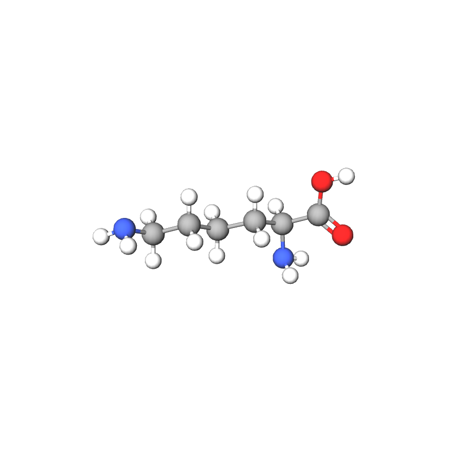
L-Lysine (abbreviated as Lys or K) is an alpha-amino acid with the chemical formula HO2CCH(NH2)(CH2)4NH2. This amino acid is an essential amino acid, which means that humans cannot synthesize it. Its codons are AAA and AAG. L-Lysine is a base, as are arginine and histidine. The epsilon-amino group often participates in hydrogen bonding and as a general base in catalysis. Common posttranslational modifications include methylation of the epsilon-amino group, giving methyl-, dimethyl-, and trimethyllysine. The latter occurs in calmodulin. Other posttranslational modifications include acetylation. Collagen contains hydroxylysine which is derived from lysine by lysyl hydroxylase. O-Glycosylation of lysine residues in the endoplasmic reticulum or Golgi apparatus is used to mark certain proteins for secretion from the cell.L-lysine is an essential amino acid. Normal requirements for lysine have been found to be about 8 g per day or 12 mg/kg in adults. Children and infants need more- 44 mg/kg per day for an eleven to-twelve-year old, and 97 mg/kg per day for three-to six-month old. Lysine is highly concentrated in muscle compared to most other amino acids. Lysine is high in foods such as wheat germ, cottage cheese and chicken. Of meat products, wild game and pork have the highest concentration of lysine. Fruits and vegetables contain little lysine, except avocados. Normal lysine metabolism is dependent upon many nutrients including niacin, vitamin B6, riboflavin, vitamin C, glutamic acid and iron. Excess arginine antagonizes lysine. Several inborn errors of lysine metabolism are known. Most are marked by mental retardation with occasional diverse symptoms such as absence of secondary sex characteristics, undescended testes, abnormal facial structure, anemia, obesity, enlarged liver and spleen, and eye muscle imbalance. Lysine also may be a useful adjunct in the treatment of osteoporosis. Although high protein diets result in loss of large amounts of calcium in urine, so does lysine deficiency. Lysine may be an adjunct therapy because it reduces calcium losses in urine. Lysine deficiency also may result in immunodeficiency. Requirements for this amino acid are probably increased by stress. Lysine toxicity has not occurred with oral doses in humans. Lysine dosages are presently too small and may fail to reach the concentrations necessary to prove potential therapeutic applications. Lysine metabolites, amino caproic acid and carnitine have already shown their therapeutic potential. Thirty grams daily of amino caproic acid has been used as an initial daily dose in treating blood clotting disorders, indicating that the proper doses of lysine, its precursor, have yet to be used in medicine. Low lysine levels have been found in patients with Parkinson's, hypothyroidism, kidney disease, asthma and depression. The exact significance of these levels is unclear, yet lysine therapy can normalize the level and has been associated with improvement of some patients with these conditions. Abnormally elevated hydroxylysines have been found in virtually all chronic degenerative diseases and coumadin therapy. The levels of this stress marker may be improved by high doses of vitamin C. Lysine is particularly useful in therapy for marasmus (wasting) and herpes simplex. It stops the growth of herpes simplex in culture, and has helped to reduce the number and occurrence of cold sores in clinical studies. Dosing has not been adequately studied, but beneficial clinical effects occur in doses ranging from 100 mg to 4 g a day. Higher doses may also be useful, and toxicity has not been reported in doses as high as 8 g per day. Diets high in lysine and low in arginine can be useful in the prevention and treatment of herpes. Some researchers think herpes simplex virus is involved in many other diseases related to cranial nerves such as migraines, Bell's palsy and Meniere's disease. Herpes blister fluid will produce fatal encephalitis in the rabbit.


Wikipedia
Lysine (symbol Lys or K) is an α-amino acid that is a precursor to many proteins. It contains an α-amino group (which is in the protonated −NH+3 form when dissolved in water), an α-carboxylic acid group (which is in the deprotonated −COO form when dissolved in water), and a side chain lysyl ((CH2)4NH2), classifying it as a basic, charged (at physiological pH), aliphatic amino acid. It is encoded by the codons AAA and AAG. Like almost all other amino acids, the α-carbon is chiral and lysine may refer to either enantiomer or a racemic mixture of both. For the purpose of this article, lysine will refer to the biologically active enantiomer L-lysine, where the α-carbon is in the S configuration.

HMDB

Lysine (Lys), also known as L-lysine is an alpha-amino acid. These are amino acids in which the amino group is attached to the carbon atom immediately adjacent to the carboxylate group (alpha carbon). Amino acids are organic compounds that contain amino (-NH2) and carboxyl (-COOH) functional groups, along with a side chain (R group) specific to each amino acid. Lysine is one of 20 proteinogenic amino acids, i.e., the amino acids used in the biosynthesis of proteins. Lysine is found in all organisms ranging from bacteria to plants to animals. It is classified as an aliphatic, positively charged or basic amino acid. In humans, lysine is an essential amino acid, meaning the body cannot synthesize it, and it must be obtained from the diet. Lysine is high in foods such as wheat germ, cottage cheese and chicken. Of meat products, wild game and pork have the highest concentration of lysine. Fruits and vegetables contain little lysine, except avocados. Normal requirements for lysine have been found to be about 8 g per day or 12 mg/kg in adults. Children and infants need more, 44 mg/kg per day for an eleven to-twelve-year old, and 97 mg/kg per day for three-to six-month old. In organisms that synthesise lysine, it has two main biosynthetic pathways, the diaminopimelate and alpha-aminoadipate pathways, which employ distinct enzymes and substrates and are found in diverse organisms. Lysine catabolism occurs through one of several pathways, the most common of which is the saccharopine pathway. Lysine plays several roles in humans, most importantly proteinogenesis, but also in the crosslinking of collagen polypeptides, uptake of essential mineral nutrients, and in the production of carnitine, which is key in fatty acid metabolism. Lysine is also often involved in histone modifications, and thus, impacts the epigenome. Lysine is highly concentrated in muscle compared to most other amino acids. Normal lysine metabolism is dependent upon many nutrients including niacin, vitamin B6, riboflavin, vitamin C, glutamic acid and iron. Excess arginine antagonizes lysine. Several inborn errors of lysine metabolism are known, such as cystinuria, hyperdibasic aminoaciduria I, lysinuric protein intolerance, propionic acidemia, and tyrosinemia I. Most are marked by mental retardation with occasional diverse symptoms such as absence of secondary sex characteristics, undescended testes, abnormal facial structure, anemia, obesity, enlarged liver and spleen, and eye muscle imbalance. Lysine also may be a useful adjunct in the treatment of osteoporosis. Although high protein diets result in loss of large amounts of calcium in urine, so does lysine deficiency. Lysine may be an adjunct therapy because it reduces calcium losses in urine. Lysine deficiency also may result in immunodeficiency. Requirements for lysine are probably increased by stress. Lysine toxicity has not occurred with oral doses in humans. Lysine dosages are presently too small and may fail to reach the concentrations necessary to prove potential therapeutic applications. Lysine metabolites, amino caproic acid and carnitine have already shown their therapeutic potential. Thirty grams daily of amino caproic acid has been used as an initial daily dose in treating blood clotting disorders, indicating that the proper doses of lysine, its precursor, have yet to be used in medicine. Low lysine levels have been found in patients with Parkinson's, hypothyroidism, kidney disease, asthma and depression. The exact significance of these levels is unclear, yet lysine therapy can normalize the level and has been associated with improvement of some patients with these conditions. Abnormally elevated hydroxylysines have been found in virtually all chronic degenerative diseases and those treated with coumadin therapy. The levels of this stress marker may be improved by high doses of vitamin C. Lysine is particularly useful in therapy for marasmus (wasting) (http://www.dcnutrition.com). Lysine has also been shown to play a role in anaemia, as lysine is suspected to have an effect on the uptake of iron and, subsequently, the concentration of ferritin in blood plasma.

Lysine 2DLysine 3D

Synonyms
Synonyms
L-lysine(S)-Lysine
lysineAminutrin
56-87-1(2S)-2,6-diaminohexanoic acid
lysine acidalpha-Lysine
h-Lys-ohL-(+)-Lysine

Identifiers
Manual Xrefs
PubChem CID5962
ChEBICHEBI:18019
ChEMBLCHEMBL8085
DrugBankDB00123
Toxic Exposome DatabaseT3D4248
MetaCycLysine
WikipediaLysine
HMDBHMDB0000182
KEGG CompoundC00047
UNIIK3Z4F929H6

Related Amino acids
Related Amino Acids

Related Amino Acids based on properties with scores

arginine [5] histidine [4] serine [3] threonine [3] tyrosine [3]
Biological activity
1. Function
Biochemical and nutritional research, pharmaceuticals, culture media, fortification of foods and feed (wheat flour), nutrient and dietary supplement, animal feed additive
2. Function
Enrichment of cereals and feeds.
3. Function
Energy fuel: Eventually, most of the ingested Lys is completely metabolized. The daily rate of Lys oxidation in healthy adults with low to moderate intake is about 27mg/kg body weight (El-Khoury et al., 2000). Lys is a particularly important energy fuel for muscles. Complete oxidation provides 4.92 kcal/g (May and Hill, 1990) and depends on adequate supplies of thiamin, riboflavin, niacin, vitamin B6, pantothenate, lipoate, ubiquinone, iron, and magnesium.
4. Function
Protein and peptide synthesis: Lys is a regular component of most proteins and many peptides. Posttranslational reactions can modify the amino group that is not engaged by the peptide bond in proteins. An important example is the conjugation of specific Lys residues in a few proteins to biotin by biotin-[propionyl-CoA-carboxylase (ATP-hydrolyzing)] ligase (EC6.3.4.10). Another type of posttranslational modification involving Lys residues consists of hydroxylation and the subsequent formation of cross-links in collagens. First, procollagen-lysine 5-dioxygenase (EC1.14.11.4) attached to the rough endoplasmic reticulum uses alpha-ketoglutarate and oxygen to hydroxylate Lys residues adjacent to glycines in procollagen. Ascorbate keeps iron in this ferroenzyme in a reduced state. Several genetically distinct isoforms exist. As procollagen extrudes into the extracellular space and forms the typical triple helix arrangements, the copper-enzyme lysyl oxidase (protein-lysine 6-oxidase, EC1.4.3.13) links strands through the formation of bonds between Lys and hydroxylysine residues. Another example is the methylation of specific Lys residues in histories, which is critical to maintaining certain chromatin segments in the inactive state (Peters et al., 2002)
5. Function
Carnitine synthesis: Lys is the critical precursor for endogenous carnitine synthesis in the liver, kidneys, and some other tissues. Daily production is about 0.2mg/kg and depends on the adequate availability of niacin, vitamin B6, folate, ascorbate, SAM, and iron. Specific lysine residues of myosin, actin, histones, and a few other proteins are trimethylated. Hydrolysis of these proteins during normal tissue turnover releases trimethyllysine, and this is hydroxylated and modified further to finally yield carnitine
6. Function
Polyamine synthesis: The PLP-containing ornithine decarboxylase (EC4.1.1.17) converts Lys into cadaverine, which plays an important role in intracellular and intercellular signaling. One example of its functions is the ability of cadaverine to prevent the escape of Shigella flexneri from phagolysosomes by blocking transepithelial signaling to polymorphonuclear cells (Fernandez et al., 2001).
References
1. Kohlmeier, M. (2015). Nutrient metabolism: Structures, functions, and genes (2nd Edition). Academic Press. 10.1016/C2010-0-64980-7
2. O'Neil, M.J. (ed.). The Merck Index - An Encyclopedia of Chemicals, Drugs, and Biologicals. Whitehouse Station, NJ: Merck and Co., Inc., 2006., p. 977 / (HSDB)
3. Lewis, R.J. Sr.; Hawley's Condensed Chemical Dictionary 15th Edition. John Wiley & Sons, Inc. New York, NY 2007., p. 773. / (HSDB)
1. Biosynthesis: Lysine Biosynthesis from L-aspartic acid
Lysine is biosynthesized from L-aspartic acid. L-Aspartic acid can be incorporated into the cell through various methods: C 4 dicarboxylate/orotate:H + symporter, glutamate/aspartate: H + symporter GltP, dicarboxylate transporter, C 4 dicarboxylate/C 4 monocarboxylate transporter DauA, and glutamate/aspartate ABC transporter. L-Aspartic acid is phosphorylated by an ATP-driven aspartate kinase resulting in ADP and L-aspartyl-4-phosphate. L-Aspartyl-4-phosphate is then dehydrogenated through an NADPH-driven aspartate semialdehyde dehydrogenase resulting in a release of phosphate, NADP, and L-aspartic 4-semialdehyde (involved in methionine biosynthesis). L-Aspartic 4-semialdehyde interacts with a pyruvic acid through a 4-hydroxy-tetrahydrodipicolinate synthase resulting in a release of hydrogen ion, water, and (2S,4S)-4-hydroxy-2,3,4,5-tetrahydrodipicolinate. The latter compound is then reduced by an NADPH-driven 4-hydroxy-tetrahydrodipicolinate reductase resulting in a release of water, NADP, and (S)-2,3,4,5-tetrahydrodipicolinate, This compound interacts with succinyl-CoA and water through a tetrahydrodipicolinate succinylase resulting in a release of coenzyme A and N-succinyl-2-amino-6-ketopimelate. This compound interacts with L-glutamic acid through an N-succinyldiaminopimelate aminotransferase resulting in oxoglutaric acid and N-succinyl-L,L-2,6-diaminopimelate. The latter compound is then desuccinylated by reacting with water through an N-succinyl-L-diaminopimelate desuccinylase resulting in a succinic acid and L,L-diaminopimelate. This compound is then isomerized through a diaminopimelate epimerase resulting in a meso-diaminopimelate (involved in peptidoglycan biosynthesis I). This compound is then decarboxylated by a diaminopimelate decarboxylase resulting in a release of carbon dioxide and L-lysine. L-Lysine is then incorporated into the lysine degradation pathway. Lysine also regulates its own biosynthesis by repressing dihydrodipicolinate synthase and also by repressing lysine-sensitive aspartokinase 3. Diaminopielate is a precursor for lysine as well as other cell wall components. Synthesis of lysine starts by converting L-aspartic acid (L-aspartate) to L-Aspartyl-4-phosphate by aspartate kinase. L-Aspartyl-4-phosphate transforms to form L-aspartic 4-semialdehyde (L-aspartate semialdehyde) by aspartate semialdehyde dehydrogenase with NADPH. L-aspartic 4-semialdehyde can start the metabolic pathway of synthesis of methionine as well as synthesis of threonine. Aspartate kinase can be regulated by its end product: L-Lysine. (http://pathbank.org/view/SMP 0000820 )

Aspartate Incorporation

L-Aspartic acid can be incorporated into the cell through various transporters such as C4 dicarboxylate/orotate:H+ symporter, glutamate/aspartate:H+ symporter GltP, dicarboxylate transporter, C4 dicarboxylate/C4 monocarboxylate transporter DauA, and glutamate/aspartate ABC transporter.

N/A

Aspartate Phosphorylation

L-Aspartic acid is phosphorylated by an ATP-driven aspartate kinase, resulting in ADP and L-aspartyl-4-phosphate.

L-Aspartic acid + ATP → L-Aspartyl-4-phosphate + ADP

L-Aspartyl-4-phosphate Dehydrogenation

L-Aspartyl-4-phosphate is dehydrogenated by an NADPH-driven aspartate semialdehyde dehydrogenase, resulting in the release of phosphate, NADP, and L-aspartic 4-semialdehyde.

L-Aspartyl-4-phosphate + NADPH → L-Aspartic 4-semialdehyde + NADP + Phosphate

Formation of (2S,4S)-4-Hydroxy-2,3,4,5-tetrahydrodipicolinate

L-Aspartic 4-semialdehyde interacts with pyruvic acid through 4-hydroxy-tetrahydrodipicolinate synthase, resulting in the release of hydrogen ion, water, and (2S,4S)-4-hydroxy-2,3,4,5-tetrahydrodipicolinate.

L-Aspartic 4-semialdehyde + Pyruvic acid → (2S,4S)-4-Hydroxy-2,3,4,5-tetrahydrodipicolinate + H + + H 2 O

Reduction of (2S,4S)-4-Hydroxy-2,3,4,5-tetrahydrodipicolinate

(2S,4S)-4-Hydroxy-2,3,4,5-tetrahydrodipicolinate is reduced by an NADPH-driven 4-hydroxy-tetrahydrodipicolinate reductase, resulting in the release of water, NADP, and (S)-2,3,4,5-tetrahydrodipicolinate.

(2S,4S)-4-Hydroxy-2,3,4,5-tetrahydrodipicolinate + NADPH → (S)-2,3,4,5-Tetrahydrodipicolinate + NADP + H 2 O

Formation of N-Succinyl-2-amino-6-ketopimelate

(S)-2,3,4,5-Tetrahydrodipicolinate interacts with succinyl-CoA and water through tetrahydrodipicolinate succinylase, resulting in the release of coenzyme A and N-succinyl-2-amino-6-ketopimelate.

(S)-2,3,4,5-Tetrahydrodipicolinate + Succinyl-CoA + H 2 O → N-Succinyl-2-amino-6-ketopimelate + CoA

Formation of N-Succinyl-L,L-2,6-diaminopimelate

N-Succinyl-2-amino-6-ketopimelate interacts with L-glutamic acid through N-succinyldiaminopimelate aminotransferase, resulting in oxoglutaric acid and N-succinyl-L,L-2,6-diaminopimelate.

N-Succinyl-2-amino-6-ketopimelate + L-Glutamic acid → N-Succinyl-L,L-2,6-diaminopimelate + Oxoglutaric acid

Formation of L,L-diaminopimelate

N-Succinyl-L,L-2,6-diaminopimelate is desuccinylated by reacting with water through N-succinyl-L-diaminopimelate desuccinylase, resulting in succinic acid and L,L-diaminopimelate.

N-Succinyl-L,L-2,6-diaminopimelate + H 2 O → L,L-Diaminopimelate + Succinic acid

Formation of meso-Diaminopimelate

L,L-Diaminopimelate is isomerized through diaminopimelate epimerase, resulting in meso-diaminopimelate.

L,L-Diaminopimelate → meso-Diaminopimelate

Formation of L-Lysine

meso-Diaminopimelate is decarboxylated by diaminopimelate decarboxylase, resulting in the release of carbon dioxide and L-lysine.

meso-Diaminopimelate + H + → L-Lysine + CO 2

1. Industrial manufacturing
Isolation from acid-hydrolyzed proteins (casein, fibrin or blood corpuscle paste
2. Industrial manufacturing
The usual raw material for the fermentation production of L-lysine is molasses...high performance microbes belong to the species Corynebacterium glutamicum or Brevibactrium lactofermentum
3. Industrial manufacturing
Extraction of natural proteins, synthetically by fermentation of glucose or other carbohydrates and by synthesis from caprolactum
Sources
Dietary Sources
Commercial Sources

Can be produced commercially through enzymatic or chemical synthesis methods

Metabolism

Kohlmeier, M. (2015)

Human Metabolism

Lys catabolism transfers the two amino groups to alpha-ketoglutarate and generates two molecules of acetyl-CoA (Figure 8.54). The main pathway proceeds via saccharopine to alpha-ketoadipate in liver cytosol, and then continues in mitochondria to acetyl-CoA. An alternative peroxisomal pathway via l-pipecolate is most important in the brain and also contributes to some extent to Lys breakdown in other extrahepatic tissues. The first two steps of Lys breakdown via the main pathway use the bifunctional protein semialdehyde synthase that combines the activities of lysine ketoglutarate reductase (EC1.5.1.8) and saccharopine dehydrogenase (EC1.5.1.9). In the end, these activities move the epsilon amino group from Lys to alpha-ketoglutarate. Oxidation by aminoadipate-semialdehyde dehydrogenase (EC1.2.1.31, magnesium-dependent) and PLP-dependent transamination by 2-aminoadipate aminotransferase (EC2.6.1.39) generate alphaketoadipate. The mitochondrial oxodicarboxylate carrier (ODC, SLC25A21) moves this intermediate from cytosol into mitochondria (Fiermonte et al., 2001), where oxidative decarboxylation by oxoglutarate dehydrogenase (EC1.2.4.2) continues its metabolism. This multisubunit enzyme contains thiamin pyrophosphate and lipoamide as covalently bound cofactors. Glutaryl-CoA dehydrogenase (EC1.3.99.7) then catalyzes both FAD-dependent oxidation and decarboxylation to crotonyl-CoA. Beta-oxidation of crotonyl-CoA finally releases two acety1-CoA molecules. Alpha-deamination by l-lysine oxidase (EC1.4.3.14) starts the pipecolate pathway of Lys breakdown. Two as-yet-uncharacterized steps then generate pipecolate. This Lys intermediate is transported into brain neurons via the high-affinity proline transporter (PROT) and may influence excitation (Galli et al., 1999). Oxidation by the FAD-containing l-pipecolate oxidase (EC1.5.3.7; IJlst et al., 2000) and nonenzymic hydration generate alpha-aminoadipate semialdehyde and thus rejoin the main pathway

L-Lysine + α-Ketoglutarate + NADPH + H + ⇌ Saccharopine + NADP +
L-Lysine and α-ketoglutarate are converted to saccharopine by the bifunctional enzyme lysine-ketoglutarate reductase/saccharopine dehydrogenase (EC1.5.1.8 and EC1.5.1.9) in the liver cytosol, using NADPH as a cofactor.
Saccharopine + NAD + + H 2 O ⇌ α-Aminoadipate-δ-semialdehyde + L-Glutamate + NADH + H +
Saccharopine is oxidized by the saccharopine dehydrogenase activity of the bifunctional enzyme (EC1.5.1.9) to form α-aminoadipate-δ-semialdehyde and L-glutamate, using NAD+ as a cofactor.
α-Aminoadipate-δ-semialdehyde + NAD + + H 2 O ⇌ α-Aminoadipate + NADH + H +
α-Aminoadipate-δ-semialdehyde is oxidized by aminoadipate-semialdehyde dehydrogenase (EC1.2.1.31) to form α-aminoadipate, using NAD+ as a cofactor. This enzyme requires magnesium.
α-Aminoadipate + α-Ketoglutarate ⇌ α-Ketoadipate + L-Glutamate
α-Aminoadipate undergoes transamination with α-ketoglutarate, catalyzed by 2-aminoadipate aminotransferase (EC2.6.1.39) using PLP as a cofactor, to produce α-ketoadipate and L-glutamate.
α-Ketoadipate + CoA + NAD + ⇌ Glutaryl-CoA + CO 2 + NADH + H +
α-Ketoadipate is transported into mitochondria by the oxodicarboxylate carrier (SLC25A21) and undergoes oxidative decarboxylation by the oxoglutarate dehydrogenase complex (EC1.2.4.2), which contains thiamin pyrophosphate and lipoamide as cofactors, to form glutaryl-CoA, CO2, and NADH.
Glutaryl-CoA + FAD ⇌ Crotonyl-CoA + CO 2 + FADH 2
Glutaryl-CoA undergoes FAD-dependent oxidation and decarboxylation by glutaryl-CoA dehydrogenase (EC1.3.99.7) to form crotonyl-CoA, CO2, and FADH2.
Crotonyl-CoA + 2 CoA + 2 NAD + + FAD + H 2 O ⇌ 2 Acetyl-CoA + 2 NADH + 2 H + + FADH 2
Crotonyl-CoA undergoes β-oxidation to release two molecules of acetyl-CoA, generating NADH and FADH2 in the process.
L-Lysine + O 2 + H 2 O ⇌ α-Keto-ε-aminocaproate + NH 3 + H 2 O 2
In the pipecolate pathway, L-lysine undergoes α-deamination by L-lysine oxidase (EC1.4.3.14) to form α-keto-ε-aminocaproate, ammonia, and hydrogen peroxide.
α-Keto-ε-aminocaproate ⇌ Pipecolate
α-Keto-ε-aminocaproate is converted to pipecolate through two uncharacterized steps in the pipecolate pathway.
Pipecolate + O 2 ⇌ Δ1-Piperideine-6-carboxylate + H 2 O 2
Pipecolate is oxidized by the FAD-containing L-pipecolate oxidase (EC1.5.3.7) to form Δ1-piperideine-6-carboxylate and hydrogen peroxide.
Δ1-Piperideine-6-carboxylate + H 2 O ⇌ α-Aminoadipate-δ-semialdehyde
Δ1-Piperideine-6-carboxylate undergoes nonenzymatic hydration to form α-aminoadipate-δ-semialdehyde, rejoining the main pathway of lysine catabolism.

Escherichia coli

The degradation of L-lysine happens in liver and it is consisted of seven reactions. L-Lysine is imported into liver through low affinity cationic amino acid transporter 2 (cationic amino acid transporter 2/SLC7A2). Afterwards, L-lysine is imported into mitochondria via mitochondrial ornithine transporter 2. L-Lysine can also be obtained from biotin metabolism. L-Lysine and oxoglutaric acid will be combined to form saccharopine by facilitation of mitochondrial alpha-aminoadipic semialdehyde synthase, and then, mitochondrial alpha-aminoadipic semialdehyde synthase will further breaks saccharopine down to allysine and glutamic acid. Allysine will be degraded to form aminoadipic acid through alpha-aminoadipic semialdehyde dehydrogenase. Oxoadipic acid is formed from catalyzation of mitochondrial kynurenine/alpha-aminoadipate aminotransferase on aminoadipic acid. Oxoadipic acid will be further catalyzed to form glutaryl-CoA, and glutaryl-CoA converts to crotonoyl-CoA, and crotonoyl-CoA transformed to 3-hydroxybutyryl-CoA. 3-Hydroxybutyryl-CoA will form Acetyl-CoA as the final product through the intermediate compound: acetoacetyl-CoA. Acetyl-CoA will undergo citric acid cycle metabolism. Carnitine is another key byproduct of lysine metabolism (not shown in this pathway).

Biochemical reactions
Bioactivity and Pharmacokinetics

Insures the adequate absorption of calcium; helps form collagen ( which makes up bone cartilage & connective tissues); aids in the production of antibodies, hormones & enzymes. Recent studies have shown that Lysine may be effective against herpes by improving the balance of nutrients that reduce viral growth. A deficiency may result in tiredness, inability to concentrate, irritability, bloodshot eyes, retarded growth, hair loss, anemia & reproductive problems.

Proteins of the herpes simplex virus are rich in L-arginine, and tissue culture studies indicate an enhancing effect on viral replication when the amino acid ratio of L-arginine to L-lysine is high in the tissue culture media. When the ratio of L-lysine to L-arginine is high, viral replication and the cytopathogenicity of herpes simplex virus have been found to be inhibited. L-lysine may facilitate the absorption of calcium from the small intestine.


Cellular Locations
Tissue Locations
1. Adsorption
Various enzymes from the stomach, pancreas, and intestine hydrolyze food proteins (Figure 8.53). The hydrogen ion/peptide cotransporter 1 (SLC15A1, PepT1) and, to a much lesser extent, the hydrogen ion/peptide cotransporter 2 (SLC15A2, PepT2) mediate the uptake of dipeptides and tripeptides with broad specificity. Free Lys can enter intestinal cells via the y+, sodium-independent cationic amino acid transporters 1 (CAT-1, SLC7A1) and 2 (CAT-2, SLC7A2). The sodium-dependent system Bo,+ seems to constitute only a minor transport route, possibly in the distal small intestine. Uptake can also proceed through the rBAT (SLC3A1)–anchored amino acid transporter BAT1/bo,+ (SLC7A9) at the brush border membrane, which can shuttle Lys into the enterocyte in exchange for a neutral amino acid plus a sodium ion (Chairoungdua et al., 1999). Export of Lys toward the pericapillary space uses two transporters that are anchored to the basolateral membrane by glycoprotein 4F2 (SLC3A2): both y+LAT1 (SLC7A7) and y+ (SLC7A6) move Lys into the basolateral space in exchange for a neutral amino acid and a sodium ion. Note: The conjugates Lys forms with other amino acids or sugars during the heating of foods in a browning (Maillard) reaction are not cleaved or absorbed in the small intestine. Examples of such heat-generated compounds include iysinoalanine, fructoselysine, and N epsilon-carboxymethyllysine.
1. Distribution
Blood circulation: While most Lys in blood is part of proteins, the concentration of free Lys in plasma is around 195µmol/l. Uptake into tissues proceeds mainly via various members of system y+. The glycoprotein-anchored heteroexchangers y+LAT1 and y+ contribute to a lesser extent to Lys uptake in some tissues. At least one of these transporters is also active in red blood cells. The mitochondrial ornithine transporter 1 (ORNT1, ornithine/citrulline carrier; SLC25A15) can move Lys from cytosol into mitochondria (Indiveri et al., 1999). Materno-fetal transfer: Since Lys is an essential amino acid, the fetus is fully dependent on transfer across the placenta. Several members of system y+ (CAT-1, CAT-4, and C AT-2B) mediate uptake from maternal circulation into the syntrophoblast layer. Export toward fetal circulation uses mainly the membrane-anchored heterodimer composed of y+LAT1 (SLC7A7) and glycoprotein 4F2 (SLC3A2). Lys is exchanged by the y+LAT1 transporter for a neutral amino acid and a sodium ion. BBB: While there is no doubt that circulating blood has to supply the essential nutrient Lys to the brain, knowledge about the mechanism for transfer across the BBB is limited. The 4F2-anchored exchange complex y+LAT2 is known to contribute significantly (Bröer et al., 2000). Adequacy of Lys intake influences flux into the brain (Tews et al., 1988)
1. Excretion
Losses of intact Lys are minimal due to efficient recovery both from the intestines and kidneys. In contrast, the dead-end metabolite methyllysine is not well reabsorbed, and most of it is excreted with urine. Nearly 5g of Lys passes across the renal glomeruli, and most of it is reabsorbed from the proximal tubular lumen. Uptake proceeds via the sodium-dependent system B°, the sodium-independent system y+ (CAT-1, SLC7A1) and 2 (CAT-2, SLC7A2), and the transporter heterodimer, consisting of BAT1/ bo,+ (SLC7A9) and rBAT (SLC3A1). The glycoprotein 4F2-1inked transporters y+LAT1 (SLC7A7) and y+LAT2 (SLC7A6) on the basolateral side mediate export toward the capillaries (Bröer et al., 2000; Bode, 2001)
1. Regulation
Lys homeostasis is maintained in part by changes in the rate of protein catabolism and Lys metabolic utilization, as well as oxidation as an energy fuel, but details of the responsible mechanisms remain to be elucidated. Lys catabolism increases greatly in response to intake (EI-Khoury et al., 1998).
Predicted Properties
Safety and Hazards

(a) Product. Lysine (L- and DL-forms). (b) Conditions of use. This substance is generally recognized as safe when used in accordance with good manufacturing or feeding practice.

Information pertaining to Adverse Effects in (1) Animals and (2) Humans

Targets and transporters
Annotations
Biotechnological production of l-lysine and its derivatives

l-lysine is an essential amino acid that contains various functional groups including α-amino, ω-amino, and α-carboxyl groups, exhibiting high reaction potential. The derivatization of these functional groups produces a series of value-added chemicals, such as cadaverine, glutarate, and d-lysine, that are widely applied in the chemical synthesis, cosmetics, food, and pharmaceutical industries.

Dietary L-lysine and calcium metabolism in humans

Calcium deficiency contributes to age-related bone loss; consequently, any preventive approach to osteoporosis should include dietary Ca adjustment or supplementation. The ideal Ca supplement would yield the greatest bioavailability. Studies in animals have shown that dietary supplements with certain amino acids, particularly L-lysine, can increase Ca absorption. Therefore, we examined the potential effect of this essential amino acid on Ca metabolism in humans. In one study, the acute effects of an oral Ca load (3 g as CaCl2) administered with or without 400 mg of L-lysine were compared in 15 healthy and 15 osteoporotic women. In all cases, the oral Ca load determined a progressive increase in serum total Ca and Ca2+ and a concomitant decrease in neophrogenous cAMP. As expected, a progressive increase in urinary Ca excretion was also observed, except in the L-lysine-treated healthy subjects, who exhibited a blunted calciuric response to the Ca load. In a second study, the effects of a short-term dietary supplementation with either L-lysine, L-valine, or L-tryptophan (800 mg/day) on 47Ca fraction absorption were compared in 45 osteoporotic patients. L-Lysine but not L-valine or L-tryptophan significantly increased the intestinal absorption of the mineral. Our results suggest that L-lysine can both enhance intestinal Ca absorption and improve the renal conservation of the absorbed Ca. The combined effects may contribute to a positive Ca balance, thus suggesting a potential usefulness of L-lysine supplements for both preventive and therapeutic interventions in osteoporosis.

Literatures
20002005201020152020010002000300040005000
PubMed Publication CountYearCount
Quiz
Information Sources
PubChem is the world's largest collection of freely accessible chemical information. Search chemicals by name, molecular formula, structure, and other identifiers. Find chemical and physical properties, biological activities, safety and toxicity information, patents, literature citations and more.
The DrugBank database is a unique bioinformatics and cheminformatics resource that combines detailed drug (i.e. chemical, pharmacological and pharmaceutical) data with comprehensive drug target (i.e. sequence, structure, and pathway) information.
The Toxin and Toxin Target Database (T3DB), or, soon to be referred as, the Toxic Exposome Database, is a unique bioinformatics resource that combines detailed toxin data with comprehensive toxin target information.
MeSH (Medical Subject Headings) is the U.S. National Library of Medicine's controlled vocabulary thesaurus used for indexing articles for PubMed.
Chemical Entities of Biological Interest (ChEBI) is a database and ontology of molecular entities focused on 'small' chemical compounds, that is part of the Open Biomedical Ontologies effort. The term 'molecular entity' refers to any constitutionally or isotopically distinct atom, molecule, ion, ion pair, radical, radical ion, complex, conformer, etc., identifiable as a separately distinguishable entity.
ChEMBL is a manually curated database of bioactive molecules with drug-like properties. It brings together chemical, bioactivity and genomic data to aid the translation of genomic information into effective new drugs.
KEGG is an encyclopedia of genes and genomes. Assigning functional meanings to genes and genomes both at the molecular and higher levels is the primary objective of the KEGG database project.
The Human Metabolome Database (HMDB) is a freely available electronic database containing detailed information about small molecule metabolites found in the human body.
Wikipedia is the free encyclopedia that anyone can edit. It contains a significant number of chemistry and biology-related pages and chemical content
Chem-bioinformatics software for the next generation of scientists. ChemAxon offers a range of products for compound design, chemical data management, drug discovery, chemistry R&D, and education.
PubMed is a free resource supporting the search and retrieval of biomedical and life sciences literature with the aim of improving health–both globally and personally.
Submit Content

Content Submission Form

Please fill out all fields and upload your document. Accepted file formats are PDF, DOC, and DOCX, upload max 5MB). Checkout the guidelines and review process

Created onUpdated On
2024-06-022024-06-02

Cite this Page

AminoADB (Amino Acid Database). (2025). AminoADB Compound Summary for AADB0012, Lysine. Retrieved April 6, 2025 from https://aminoadb.org/aminoacid/lysine.php.