Arginine | C6H14N4O2 | Amino Acid Database

Generic NameIDType
ArginineAADB0002Essential
The *conditionally essential amino acid l-arginine (Arg) is used for high-energy phosphate storage in muscle (phosphoarginine and creatine), protein synthesis, and nitric oxide production. It is also used as an energy fuel; its complete oxidation requires thiamin, riboflavin, niacin, vitamin B6, pantothenate, lipoate, ubiquinone, iron, and magnesium. Adequate amounts are consumed with protein from most sources. Dietary supplements with manufactured Arg are commercially available. Due to the possibility of endogenous synthesis from l-glutamate (Glu), dietary intake is not usually needed except in newborn infants. Increased dietary intake may be beneficial for wound healing, tissue repair, and immune function when needs are increased due to severe illness, infection, or injury. Prolonged lack of protein causes growth failure, loss of muscle mass, and organ damage. Excessive intake: Very high intake of protein and mixed amino acids (i.e., more than three times the RDA (recommended dietary allowance), which is 2.4g/kg) is thought to increase the risk of renal glomerular sclerosis and accelerate osteoporosis. The risk from high intake of manufactured supplements is not completely understood but may include liver and kidney failure, mental disturbances, and other severe dysfunction. (Kohlmeier 2015)
Amino acid ClassSide Chain Type
BasicPolar, positively charged
Kohlmeier, M. (2015). Nutrient metabolism: Structures, functions, and genes (2nd Edition). Academic Press. 10.1016/C2010-0-64980-7
ChEBI
ChEBI :16977

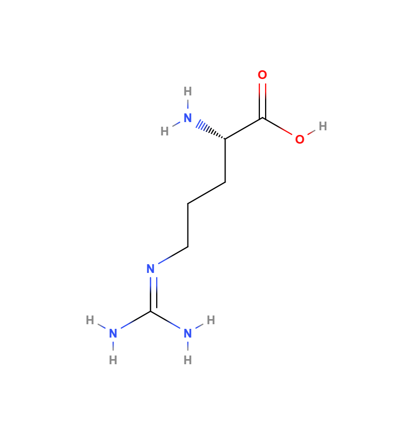
L-arginine is an L-alpha-amino acid that is the L-isomer of arginine. It has a role as a nutraceutical, a biomarker, a micronutrient, an Escherichia coli metabolite and a mouse metabolite. It is a glutamine family amino acid, a proteinogenic amino acid, an arginine and a L-alpha-amino acid. It is a conjugate base of a L-argininium(1+). It is a conjugate acid of a L-argininate. It is an enantiomer of a D-arginine.

Descriptions
DrugBank
An essential amino acid that is physiologically active in the L-form.

MeSH

1: Arginine An essential amino acid that is physiologically active in the L-form.


T3DB

Arginine is an essential amino acid that is physiologically active in the L-form. In mammals, arginine is formally classified as a semiessential or conditionally essential amino acid, depending on the developmental stage and health status of the individual. Infants are unable to effectively synthesize arginine, making it nutritionally essential for infants. Adults, however, are able to synthesize arginine in the urea cycle. Arginine can be considered to be a basic amino acid as the part of the side chain nearest to the backbone is long, carbon-containing and hydrophobic, whereas the end of the side chain is a complex guanidinium group. With a pKa of 12.48, the guanidinium group is positively charged in neutral, acidic and even most basic environments. Because of the conjugation between the double bond and the nitrogen lone pairs, the positive charge is delocalized. This group is able to form multiple H-bonds. L-arginine is an amino acid that has numerous functions in the body. It helps dispose of ammonia, is used to make compounds such as nitric oxide, creatine, L-glutamate, L-proline, and it can be converted to glucose and glycogen if needed. In large doses, L-arginine also stimulates the release of hormones growth hormone and prolactin. Arginine is a known inducer of mTOR (mammalian target of rapamycin) and is responsible for inducing protein synthesis through the mTOR pathway. mTOR inhibition by rapamycin partially reduces arginine-induced protein synthesis (A13142). Catabolic disease states such as sepsis, injury, and cancer cause an increase in arginine utilization, which can exceed normal body production, leading to arginine depletion. Arginine also activates AMP kinase (AMPK) which then stimulates skeletal muscle fatty acid oxidation and muscle glucose uptake, thereby increasing insulin secretion by pancreatic beta-cells (A13143). Arginine is found in plant and animal proteins, such as dairy products, meat, poultry, fish, and nuts. The ratio of L-arginine to lysine is also important - soy and other plant proteins have more L-arginine than animal sources of protein.


Wikipedia

Arginine is the amino acid with the formula (H2N)(HN)CN(H)(CH2)3CH(NH2)CO2H. The molecule features a guanidino group appended to a standard amino acid framework. At physiological pH, the carboxylic acid is deprotonated (−CO2) and both the amino and guanidino groups are protonated, resulting in a cation. Only the l-arginine (symbol Arg or R) enantiomer is found naturally. Arg residues are common components of proteins. It is encoded by the codons CGU, CGC, CGA, CGG, AGA, and AGG. The guanidine group in arginine is the precursor for the biosynthesis of nitric oxide. Like all amino acids, it is a white, water-soluble solid.


HMDB

L-argininium(1+), also known as L-Arginine or DL Arginine acetate, monohydrate, is classified as a member of the L-alpha-amino acids. L-alpha-amino acids are alpha amino acids which have the L-configuration of the alpha-carbon atom. L-argininium(1+) is considered to be soluble (in water) and acidic

Arginine 2DArginine 3D

Synonyms
Synonyms
L-arginine(S)-2-Amino-5-guanidinopentanoic acid
arginineL(+)-Arginine
74-79-3H-Arg-OH
L-(+)-Arginine(L)-Arginine
L-ArgArginina

Identifiers
Manual Xrefs
PubChem CID6322
ChEBICHEBI:29016
ChEMBLCHEMBL1485
DrugBankDB00125
Toxic Exposome DatabaseT3D4289
MetaCycAsparagine
WikipediaC00001341
HMDBHMDB0000168
CAS Number70-47-3
EC Number230-571-3
KEGGC02385
UNII94ZLA3W45F

Related Amino acids
Related Amino Acids
Biological activity
1. Function
An essential amino acid; Used as an ammonia detoxicant and diagnostic aid; [Merck Index] Used as a nutritional supplement for foods; (FDA) Used in biochemical research, pharmaceuticals, as a dietary supplement, and medication; (HSDB)
2. Function
Used for nutritional supplementation, also for treating dietary shortage or imbalance. (T3D4289)
3. Function
Energy fuel: Most catabolic pathways of Arg lead to complete oxidation with an energy yield of 3.3kcal/g (May and Hill, 1990). The necessary reactions depend on thiamin, riboflavin, niacin, vitamin B6, pantothenate, lipoate, ubiquinone, iron, and magnesium
4. Function
Protein synthesis: Significant amounts of Arg are needed for protein synthesis. Arginine-tRNA ligase (EC6.1.1.19) loads Arg onto its specific tRNA. Hair protein has a relatively high Arg content and is adversely affected by Arg deficiency
5. Function
Agmatin: The amine (Figure 8.77) produced through the mitochondrial decarboxylation of Arg (arginine decarboxylase, EC4.1.1.19, PLP-dependent) interacts with neurotransmitter receptors in the brain, is an acceptor of ADP-ribose (immunemodulator), tempers the proliferative effects of polyamines (through its effects on ornithine decarboxylase and the putrescine transporter), causes vasodilatation and increases the renal glomerular filtration rate (GFR), and slows nitric oxide production (Blantz et al., 2000).
6. Function
Nitric oxide synthesis: All isoforms (NOS1, NOS2, and NOS3) of nitric oxide synthase (EC1.14.13.39) produce nitric oxide from Arg (Figure 8.78). They require 1 mole of BH4 and 1 mole of heme per dimer as cofactors (Rafferty et al., 1999). Diminished BH4 synthesis and the resulting decrease in nitric oxide production has been suggested to contribute importantly to impaired angiogenesis (Marinos et al., 2001), epithelial dysfunction, and insufficient vasodilation (Gruhn et al., 2001). Nitric oxide synthesis is inhibited by ADMA at moderately elevated concentrations, but not by symmetric N(G),N′(G)-dimethylarginine (Masuda et al., 2002). Plasma concentrations of both compounds are below 0.5µmol/l in normal subjects (Teerlink et al., 2002) but distinctly elevated in people with diabetes, renal failure, and other diseases
7. Function
High-energy phosphates: Daily production of the high-energy storage compound creatine is about 15mg/kg, using Arg and glycine as precursors. The synthesis takes place in two stages, starting in the kidneys, and coming to completion in the liver. Another functionally related energy-storage compound for muscle is phosphoarginine
8. Function
Hormone stimulation: Increased Arg intake elicits the release of prolactine, insulin, glucagon, growth hormone, and enhances the number and responsiveness of circulating lymphocytes to mitogens. Arginine deiminase may inhibit the proliferation of human leukemia cells (by inducing cell growth arrest in the G1- and/or S-phase and apoptosis) more potently than analogous asparaginase treatment (Gong et al., 2000)
References
1. Kohlmeier, M. (2015). Nutrient metabolism: Structures, functions, and genes (2nd Edition). Academic Press. 10.1016/C2010-0-64980-7
2. Merck Index - O'Neil MJ, Heckelman PE, Dobbelaar PH, Roman KJ (eds). The Merck Index, An Encyclopedia of Chemicals, Drugs, and Biologicals, 15th Ed. Cambridge, UK: The Royal Society of Chemistry, 2013. / (HSDB)
3. Toxin and Toxin Target Database (T3D4289)
1. Biosynthesis: Biosynthesis via the linear pathway in E. coli.
Arginine biosynthesis via the linear pathway in E. coli. The linear pathway is a metabolic route for the biosynthesis of arginine, which occurs in organisms like E. coli. This pathway involves a series of enzymatic reactions that convert glutamate to arginine. Five steps involving N-acetylated intermediates lead to ornithine, and three additional steps are required to convert ornithine into arginine.

N-acetylglutamate synthesis

L-glutamate is acetylated by the enzyme N-acetylglutamate synthase (NAGS, argA) to form N-acetyl-L-glutamate.

L-glutamate + acetyl-CoA ⇌ N-acetyl-L-glutamate + CoASH

N-acetylglutamate phosphorylation

N-acetyl-L-glutamate is phosphorylated by the enzyme N-acetylglutamate kinase (NAGK, argB) to form N-acetyl-L-glutamyl-5-phosphate.

N-acetyl-L-glutamate + ATP ⇌ N-acetyl-L-glutamyl-5-phosphate + ADP

N-acetylglutamyl-5-semialdehyde formation

N-acetyl-γ-glutamylphosphate reductase (argC) catalyzes the reduction of N-acetyl-L-glutamyl-5-phosphate to N-acetyl-L-glutamyl-5-semialdehyde.

N-acetyl-L-glutamyl-5-phosphate + NADPH + H⁺ ⇌ N-acetyl-L-glutamyl-5-semialdehyde + NADP⁺ + Pi

N-acetylornithine synthesis

N-acetylornithine aminotransferase (argD) catalyzes the transfer of an amino group from glutamate to N-acetyl-L-glutamyl-5-semialdehyde, forming N-acetyl-L-ornithine and α-ketoglutarate.

N-acetyl-L-glutamyl-5-semialdehyde + glutamate ⇌ N-acetyl-L-ornithine + α-ketoglutarate

Ornithine synthesis

N-acetylornithine deacetylase (AO, argE) removes the acetyl group from N-acetyl-L-ornithine, producing L-ornithine and acetate.

N-acetyl-L-ornithine + H₂O ⇌ L-ornithine + acetate

Citrulline synthesis

Ornithine carbamoyltransferase (OTC, argF/argI) catalyzes the condensation of L-ornithine with carbamoyl phosphate, forming L-citrulline and inorganic phosphate.

L-ornithine + carbamoyl phosphate ⇌ L-citrulline + Pi

Argininosuccinate synthesis

Argininosuccinate synthetase (argG) catalyzes the condensation of L-citrulline with aspartate to form L-argininosuccinate.

L-citrulline + ATP + L-aspartate ⇌ L-argininosuccinate + AMP + PPi

Arginine synthesis

Argininosuccinase (argH) cleaves L-argininosuccinate to produce L-arginine and fumarate.

L-argininosuccinate ⇌ L-arginine + fumarate

1. Industrial manufacturing
It is precipitated as the flavinate from gelatin hydrolyzate in industry.
2. Industrial manufacturing
An essential amino acid for human development; precursor for nitric oxide; Isolation from etiolated lupine seedlings: E. Schulze, E. Steiger, Ber 19, 1177 (1886)
Sources
Dietary Sources
Natural Sources
Commercial Sources

Arginine can be produced commercially through microbial fermentation, enzymatic synthesis, or chemical synthesis methods.

Metabolism

Kohlmeier, M. (2015)

Human Metabolism

Arg breakdown in the liver proceeds mainly via ornithine and glutamate to alpha-ketoglutarate and releases four nitrogens (two with urea and another two in transamination reactions). Several of the necessary steps are the same used for Arg synthesis operating in the reverse direction (Figure 8.76). Complete oxidation requires adequate supplies of thiamin, riboflavin, vitamin B6, niacin, pantothenate, lipoate, ubiquinone, iron, and magnesium. The first step of Arg catabolism (in cytosol) uses the final enzyme of urea synthesis, the manganese-dependent arginase (EC3.5.3.1). Transport of the resulting ornithine into mitochondria by the ornithine/citrulline carrier (SLC25A15) is the rate-limiting step of Arg catabolism. This transporter usually exchanges an inwardly carried ornithine molecule for an outwardly transported citrulline molecule. Since a proton instead of citrulline can also serve as the counter ion (Indiveri et al., 1999), removal of ornithine from the urea cycle sequence does not impede the functioning of the ornithine/citrulline carrier. The delta-amino group of ornithine can then be moved by mitochondrial OAT (ornithine transaminase, EC2.6.1.13, PLP-dependent) to alpha-ketoglutarate, pyruvate, or glyoxylate. Glutamate-5-semialdehyde dehydrogenase (EC1.2.1.41) produces glutamate, which can then be transaminated by more than a dozen PLP-dependent aminotransferases, including aspartate aminotransferase (EC2.6.1.1) and alanine aminotransferase (EC2.6.1.2), to the Krebs-cycle intermediate alpha-ketoglutarate. Protein arginine N-methyltransferases use SAM to methylate a small portion of arginyl residues in specific proteins. This activity is important for mRNA splicing, RNA transport, transcription control, signal transduction, and maturation of protein such as the MBP. Type I enzymes dimethylate asymmetrically, type II enzymes methylate proteins such as the MBP symmetrically. Protein–arginine N-methyltransferases 1, 3, 4, and 6 are type I enzymes, whereas isoform 5 is a type II enzyme (Frankel et al., 2002). Breakdown of symmetrically methylated proteins releases symmetric N(G),N′(G)- dimethylarginine, and breakdown of asymmetrically dimethylated proteins generates asymmetric N(G),N(G)-dimethylarginine (ADMA). Dimethylargininase (dimethylarginine dimethylaminohydrolase, EC3.5.3.18, contains zinc), which is present in two isoforms in most tissues, converts these metabolites to l-citrulline by cleaving off methylamine or dimethylamine, respectively.

L-Arginine + H 2 O → Ornithine + Urea
The first step of arginine catabolism in the cytosol uses the enzyme arginase (EC 3.5.3.1) to convert L-arginine and water into ornithine and urea.
Ornithine (cytosol) → Ornithine (mitochondria)
Ornithine is transported from the cytosol into the mitochondria by the ornithine/citrulline carrier (SLC25A15). This step is the rate-limiting step of arginine catabolism.
Ornithine + α-Ketoglutarate → Glutamate-γ-semialdehyde + Glutamate
Mitochondrial ornithine aminotransferase (OAT, EC 2.6.1.13) transfers the delta-amino group of ornithine to α-ketoglutarate, forming glutamate-γ-semialdehyde and glutamate.
Glutamate-γ-semialdehyde + NAD + + H 2 O → Glutamate + NADH + H +
Glutamate-γ-semialdehyde dehydrogenase (EC 1.2.1.41) oxidizes glutamate-γ-semialdehyde to glutamate, producing NADH and a proton in the process.
Glutamate + α-Keto acid ⇌ α-Ketoglutarate + Amino acid
Various PLP-dependent aminotransferases, including aspartate aminotransferase (EC 2.6.1.1) and alanine aminotransferase (EC 2.6.1.2), transaminate glutamate to α-ketoglutarate, forming different amino acids.
L-Glutamate + ATP → γ-Glutamyl phosphate + ADP
Glutamate kinase catalyzes the phosphorylation of L-glutamate to γ-glutamyl phosphate using ATP as the phosphate donor.
γ-Glutamyl phosphate + NAD + + Pi → α-Ketoglutarate + NADH + H 2 O
γ-Glutamyl phosphate is further oxidized to α-ketoglutarate, producing NADH and releasing phosphate and water.
Protein-arginine + SAM → Methylated protein + SAH
Protein arginine N-methyltransferases use S-adenosylmethionine (SAM) to methylate arginine residues in proteins, producing S-adenosylhomocysteine (SAH).
Symmetric/Asymmetric N(G),N′(G)-dimethylarginine + H 2 O → L-Citrulline + Methylamine/Dimethylamine
Dimethylargininase (EC 3.5.3.18) converts symmetric N(G),N′(G)-dimethylarginine or asymmetric N(G),N(G)-dimethylarginine to L-citrulline, releasing methylamine or dimethylamine, respectively.

Amino Acids Are Precursors of Creatine

Creatine is synthesized from glycine, arginine, and methionine in a two-step process that involves the transfer of an amidino group from arginine to glycine, forming guanidinoacetate, and then the methylation of guanidinoacetate by S-adenosylmethionine to form creatine. Glutathione (GSH), present in plants, animals, and some bacteria, often at high levels, can be thought of as a redox buffer. Creatine supplementation is an effective ergogenic aid to augment resistance training and improve intense, short duration, intermittent performance. (Forbes, Scott C et al. 2023). Creatine is a key player in heart contraction and energy metabolism. (Balestrino, Maurizio. 2021). Creatine, a widely available nutritional supplement, has the potential to improve these disruptions in some patients, and early clinical trials indicate that it may have efficacy as an antidepressant agent. (Kious, Brent M et al. 2019). Phosphocreatine, derived from creatine, is an important energy buffer in skeletal muscle

Glycine + Arginine → Guanidinoacetate + Ornithine
Glycine and arginine are combined by the enzyme arginine:glycine amidinotransferase (AGAT) to form guanidinoacetate and ornithine.
Guanidinoacetate + S-Adenosylmethionine → Creatine + S-Adenosylhomocysteine
Guanidinoacetate is methylated by S-adenosylmethionine in a reaction catalyzed by guanidinoacetate N-methyltransferase (GAMT), producing creatine and S-adenosylhomocysteine.
Creatine + ATP ⇌ Phosphocreatine + ADP
Creatine can reversibly accept a phosphate group from ATP to form phosphocreatine, catalyzed by creatine kinase. This reaction serves as an energy buffer in tissues with high energy demands, such as skeletal muscle.

Nitric Oxide Biosynthesis

Nitric oxide (NO) is a gaseous signaling molecule synthesized from the amino acid arginine by the enzyme nitric oxide synthase (NOS). NO plays important roles in various physiological processes, including neurotransmission, blood clotting, and regulation of blood pressure.

Arginine + 1.5 NADPH + 1.5 H⁺ + 2 O₂ → Citrulline + NO + 1.5 NADP⁺ + 2 H₂O
Nitric oxide synthase catalyzes the conversion of arginine to citrulline, with the release of nitric oxide (NO). This reaction is NADPH-dependent and requires molecular oxygen. NOS is a dimeric enzyme structurally related to NADPH cytochrome P-450 reductase. Each subunit contains one bound molecule of each of four different cofactors. The activity of NOS is stimulated by the binding of Ca²⁺-calmodulin. Cofactors: FMN, FAD, Tetrahydrobiopterin, Fe³⁺ heme

Escherichia coli

The arginine and proline metabolism pathway illustrates the biosynthesis and metabolism of several amino acids including arginine, ornithine, proline, citrulline, and glutamate in mammals. In adult mammals, the synthesis of arginine takes place primarily through the intestinal-renal axis (PMID: 19030957). In particular, the amino acid citrulline is first synthesized from several other amino acids (glutamine, glutamate, and proline) in the mitochondria of the intestinal enterocytes (PMID: 9806879). The mitochondrial synthesis of citrulline starts with the deamination of glutamine to glutamate via mitochondrial glutaminase. The resulting mitochondrial glutamate is converted into 1-pyrroline-5-carboxylate via pyrroline-5-carboxylate synthase (P5CS). Alternately, the 1-pyrroline-5-carboxylate can be generated from mitochondrial proline via proline oxidase (PO). Ornithine aminotransferase (OAT) then converts the mitochondrial 1-pyrroline-5-carboxylate into ornithine and the enzyme ornithine carbamoyltransferase (OCT -- using carbamoyl phosphate) converts the ornithine to citrulline (PMID: 19030957). After this, the mitochondrial citrulline is released from the small intestine enterocytes and into the bloodstream where it is taken up by the kidneys for arginine production. Once the citrulline enters the kidney cells, the cytosolic enzyme argininosuccinate synthetase (ASS) will combine citrulline with aspartic acid to generate argininosuccinic acid. After this step, the enzyme argininosuccinate lyase (ASL) will remove fumarate from argininosuccinic acid to generate arginine. The resulting arginine can either stay in the cytosol where it is converted to ornithine via arginase I (resulting in the production of urea) or it can be transported into the mitochondria where it is decomposed into ornithine and urea via arginase II. The resulting mitochondrial ornithine can then be acted on by the enzyme ornithine amino transferase (OAT), which combines alpha-ketoglutarate with ornithine to produce glutamate and 1-pyrroline-5-carboxylate. The mitochondrial enzyme pyrroline-5-carboxylate dehydrogenase (P5CD) acts on the resulting 1-pyrroline-5-carboxylate (using NADPH as a cofactor) to generate glutamate. Alternately, the mitochondrial 1-pyrroline-5-carboxylate can be exported into the kidney cell’s cytosol where the enzyme pyrroline-5-carboxylate reductase (P5CR) can convert it to proline. While citrulline-to-arginine production primarily occurs in the kidney, citrulline is readily converted into arginine in other cell types, including adipocytes, endothelial cells, myocytes, macrophages, and neurons. Interestingly, chickens and cats cannot produce citrulline via glutamine/glutamate due to a lack of a functional pyrroline-5-carboxylate synthase (P5CS) in their enterocytes (PMID: 19030957).

Biochemical reactions
Bioactivity and Pharmacokinetics

Studies have shown that is has improved immune responses to bacteria, viruses and tumor cells; promotes wound healing and regeneration of the liver; causes the release of growth hormones; considered crucial for optimal muscle growth and tissue repair.

Many of supplemental L-arginine's activities, including its possible anti-atherogenic actions, may be accounted for by its role as the precursor to nitric oxide or NO. NO is produced by all tissues of the body and plays very important roles in the cardiovascular system, immune system and nervous system. NO is formed from L-arginine via the enzyme nitric oxide synthase or synthetase (NOS), and the effects of NO are mainly mediated by 3,'5' -cyclic guanylate or cyclic GMP. NO activates the enzyme guanylate cyclase, which catalyzes the synthesis of cyclic GMP from guanosine triphosphate or GTP. Cyclic GMP is converted to guanylic acid via the enzyme cyclic GMP phosphodiesterase. NOS is a heme-containing enzyme with some sequences similar to cytochrome P-450 reductase. Several isoforms of NOS exist, two of which are constitutive and one of which is inducible by immunological stimuli. The constitutive NOS found in the vascular endothelium is designated eNOS and that present in the brain, spinal cord and peripheral nervous system is designated nNOS. The form of NOS induced by immunological or inflammatory stimuli is known as iNOS. iNOS may be expressed constitutively in select tissues such as lung epithelium. All the nitric oxide synthases use NADPH (reduced nicotinamide adenine dinucleotide phosphate) and oxygen (O2) as cosubstrates, as well as the cofactors FAD (flavin adenine dinucleotide), FMN (flavin mononucleotide), tetrahydrobiopterin and heme. Interestingly, ascorbic acid appears to enhance NOS activity by increasing intracellular tetrahydrobiopterin. eNOS and nNOS synthesize NO in response to an increased concentration of calcium ions or in some cases in response to calcium-independent stimuli, such as shear stress. In vitro studies of NOS indicate that the Km of the enzyme for L-arginine is in the micromolar range. The concentration of L-arginine in endothelial cells, as well as in other cells, and in plasma is in the millimolar range. What this means is that, under physiological conditions, NOS is saturated with its L-arginine substrate. In other words, L-arginine would not be expected to be rate-limiting for the enzyme, and it would not appear that supraphysiological levels of L-arginine which could occur with oral supplementation of the amino acid^would make any difference with regard to NO production. The reaction would appear to have reached its maximum level. However, in vivo studies have demonstrated that, under certain conditions, e.g. hypercholesterolemia, supplemental L-arginine could enhance endothelial-dependent vasodilation and NO production.


1. Adsorption
Arg-containing proteins are digested, as all other proteins, by an array of secreted enzymes from the stomach, pancreas, and small intestine, and by aminopeptidases at the brush border membrane (Figure 8.75). The combined action of these enzymes releases small peptides and free amino acids. Dipeptides and tripeptides are taken up through the hydrogen ion/peptide cotransporters 1 (PepT1, SLC15A1) and 2 (PepT2, SLC15A2). Sodium-dependent transport systems are known to facilitate Arg uptake into enterocytes of the distal small intestine, including at least one high-capacity, low-affinity system and a low-capacity, highaffinity system (Iannoli et al., 1998). One of these is the sodium-dependent transport system B°. Even more important is the sodium-independent uptake via system y+ and BAT1/bo,+ (SLC7A9, linked to rBAT) in exchange for a neutral amino acid plus a sodium ion. The transporters y+LAT1 (SLC7A7) and y+LAT2 (SLC7A6) have characteristics of amino acid system y+ and work only in conjunction with the same membrane glycoprotein anchor 4F2 (SLC3A2). Both mediate Arg export across the basolateral membrane in equimolar exchange for glutamine or another neutral amino acid (Bröer et al., 2000; Bode, 2001). While these systems are not driven by the sodium gradient, they transport a sodium ion together with glutamine in compensation for the charge in Arg. Enterocytes use about 40% of the dietary Arg for their own energy needs and protein synthesis. Only about 60% of the absorbed Arg reach the portal bloodstream. On the other hand, the enterocytes are a major site of de novo Arg synthesis from glutamine, glutamate, and proline (Wu and Morris, 1998).
1. Distribution
Blood circulation: Plasma concentration of Arg, typically around 94µmol/l (Teerlink et al., 2002), is lowest during the early morning hours and increases significantly after meals (Tsai and Huang, 1999). Uptake from the blood into tissues occurs mainly via sodium-independent transporters that have the characteristics of system y+ (Wu and Morris, 1998). CAT-1, CAT-2A, and CAT-2B are representatives of that family of transporters. The CAT-2 isoforms A and B are especially expressed in muscle and macrophages (Kakuda et al., 1998). The Arg-derived amine agmatine enters cells via the polyamine transporter (Satriano et al., 2001). Materno-fetal transfer: Several members of the y+ amino acid transporter family (CAT-1, CAT-4, and CAT-2B) are present at the maternal side of the syntrophoblast, but Arg uptake from maternal circulation appears to be made up of only a small proportion of total amino acid transfer (Cetin, 2001). Transfer across the basal membrane into fetal blood uses the heterodimeric complex, consisting of y+ LATI and 4F2 (SLC7A7 + SLC3A2). BBB: System y+ is the main conduit of transport for Arg into the brain. CAT-3 is a brain-specific form in rats. Additional transporters may contribute to transport in either direction.
1. Excretion
At its typical plasma concentration, more than 6g of Arg is filtered daily in a 70-kg man. Most of this is recovered from the proximal tubular lumen through the sodium-dependent system Bo,+ (SLC6A14) and the sodium-independent systems y+ and BAT1/bo,+ (SLC7A9, linked to rBAT). The mechanisms are the same as those mediating small intestinal Arg uptake. On the basolateral side, again the transporters y+LAT1 (SLC7A7) and y+LAT2 (SLC7A6) mediate export toward the capillaries (Bröer et al., 2000; Bode, 2001). The inner medullary collecting ducts in the kidneys take up Arg via CAT1 (system y+) but do not express CAT2A, CAT2B, or CAT3 (Wu et al., 2000).
1. Regulation
Information on regulatory events in Arg homeostasis is still very incomplete. The control of dietary intake as well as differential distribution to the liver and other tissues contribute to a steady supply for vital functions and the avoidance of excess. Selection of foods may be influenced by their Arg content as suggested by observations in rats (Yamamoto and Muramatsu, 1987). Subjects with citrullinemia, an inborn error of arginine synthesis, have been reported to crave beans, peas, and peanuts, which are high in arginine (Walser, 1983); this observation could be another indication of some kind of feedback control of Arg intake. Arg potentiates glucose-induced insulin secretion (Thams and Capito, 1999), which in turn shifts Arg away from use for gluconeogenesis and toward use for protein synthesis. The rate of endogenous Arg synthesis appears to be little affected by intake levels (Castillo et al., 1994). Inflammatory cytokines and endotoxin acting on system y+ transporters enhance uptake of Arg into particular tissues. This system is expressed only at a low level in hepatocytes but strongly induced by cytokines (Kakuda et al., 1998). The control of CAT-2 expression indicates the level of complexity at work. Not only does. this gene encode for two distinct isoforms (CAT-2 and CAT-2A) with 10-fold difference in substrate binding affinity, but it also has four separate fully functional promoters that allow differential response to a particular stimulant (Kakuda et al., 1998)
Predicted Properties
Safety and Hazards

(a) Product. Arginine (L- and DL-forms). (b) Conditions of use. This substance is generally recognized as safe when used in accordance with good manufacturing or feeding practice.

Information pertaining to Adverse Effects in (1) Animals and (2) Humans

Targets and transporters

No proteins available.

Annotations
L-arginine supplementation in peripheral arterial disease: no benefit and possible harm

Background: L-arginine is the precursor of endothelium-derived nitric oxide, an endogenous vasodilator. L-arginine supplementation improves vascular reactivity and functional capacity in peripheral arterial disease (PAD) in small, short-term studies. We aimed to determine the effects of long-term administration of L-arginine on vascular reactivity and functional capacity in patients with PAD. Methods and results: The Nitric Oxide in Peripheral Arterial Insufficiency (NO-PAIN) study was a randomized clinical trial of oral L-arginine (3 g/d) versus placebo for 6 months in 133 subjects with intermittent claudication due to PAD in a single-center setting. The primary end point was the change at 6 months in the absolute claudication distance as assessed by the Skinner-Gardner treadmill protocol. L-arginine supplementation significantly increased plasma L-arginine levels. However, measures of nitric oxide availability (including flow-mediated vasodilation, vascular compliance, plasma and urinary nitrogen oxides, and plasma citrulline formation) were reduced or not improved compared with placebo. Although absolute claudication distance improved in both L-arginine- and placebo-treated patients, the improvement in the L-arginine-treated group was significantly less than that in the placebo group (28.3% versus 11.5%; P=0.024). Conclusions: In patients with PAD, long-term administration of L-arginine does not increase nitric oxide synthesis or improve vascular reactivity. Furthermore, the expected placebo effect observed in studies of functional capacity was attenuated in the L-arginine-treated group. As opposed to its short-term administration, long-term administration of L-arginine is not useful in patients with intermittent claudication and PAD.

The effect of L-arginine and creatine on vascular function and homocysteine metabolism

Studies with L-arginine supplementation have shown inconsistent effects on endothelial function. The generation of guanidinoacetate (GAA) from L-arginine with subsequent formation of creatine and homocysteine and consumption of methionine may reduce the pool of L-arginine available for nitric oxide generation. Experimental studies suggest that creatine supplementation might block this pathway. We sought to determine the effects of L-arginine, creatine, or the combination on endothelium-dependent vasodilation and homocysteine metabolism in patients with coronary artery disease. Patients with coronary artery disease were randomized to L-arginine (9 g/day), creatine (21 g/day), L-arginine plus creatine, or placebo for 4 days (n = 26-29/group). Brachial artery flow-mediated dilation and plasma levels of L-arginine, creatine, homocysteine, methionine, and GAA were measured at baseline and follow-up. L-arginine and creatine supplementation had no effects on vascular function. L-arginine alone increased GAA (p < 0.01) and the ratio of homocysteine to methionine (p < 0.01), suggesting increased methylation demand. The combination of creatinine and L-arginine did not suppress GAA production or prevent the increase in homocysteine-to-methionine ratio. Unexpectedly, creatine supplementation (alone or in combination with L-arginine) was associated with an 11-20% increase in homocysteine concentration (p < 0.05), which was not attributable to worsened renal function, providing evidence against an effect of creatine on decreasing methylation demand. In conclusion, the present study provides no evidence that L-arginine supplementation improves endothelial function and suggests that l-arginine may increase methylation demand. Creatine supplementation failed to alter the actions of L-arginine on vascular function or suppress methylation demand. The unexpected increase in homocysteine levels following creatine supplementation could have adverse effects and merits further study, since creatine is a commonly used dietary supplement.

Therapeutic Benefits of l-Arginine: An Umbrella Review of Meta-analyses

Objective l-Arginine is a semi-essential amino acid that is the substrate for nitric oxide production by vascular endothelial and immune cells. Nitric oxide production by these cells is essential for both blood pressure regulation and immune regulation. However, there is much discrepancy in the literature when it comes to randomized controlled studies, and so this umbrella review of published meta-analyses was performed to examine the efficacy of l-arginine’s role as a therapeutic agent. Methods There was an overall search of the literature from January 1, 1980 through December 31, 2015 of three separate databases—PubMed, Cochrane Library, and Cumulative Index to Nursing and Allied Health Literature—using the following search strategy: (arginine) AND (meta-analysis OR systematic review). Only English language publications were retrieved that provided quantitative statistical analysis of outcomes on blood pressure and immune function. Results The 7 meta-analyses that were included in this umbrella review reported significant positive benefits for reducing systolic and diastolic blood pressure in hypertensive adults by 2.2 to 5.4 mm Hg and 2.7 to 3.1 mm Hg, respectively, reducing diastolic blood pressure in pregnant women with gestational hypertension by 4.9 mm Hg, and reducing the length of stay in the hospital for surgical patients; in addition, 2 of the 3 meta-analyses indicated a 40% reduction in the incidence of hospital-acquired infections. However, these positive results should be considered with caution because statistically significant heterogeneity was observed in 5 of the 7 meta-analyses. Conclusions Some evidence appears to support the benefit of l-arginine supplementation for reducing systolic and diastolic blood pressure in hypertensive adults and reducing the incidence of hospital-acquired infections and the length of stay in the hospital for surgical patients. Given the limitations of the included studies, interpretations should be made with caution.

Literatures
2000200520102015202001000200030004000
PubMed Publication CountYearCount
Quiz
Information Sources
PubChem is the world's largest collection of freely accessible chemical information. Search chemicals by name, molecular formula, structure, and other identifiers. Find chemical and physical properties, biological activities, safety and toxicity information, patents, literature citations and more.
The DrugBank database is a unique bioinformatics and cheminformatics resource that combines detailed drug (i.e. chemical, pharmacological and pharmaceutical) data with comprehensive drug target (i.e. sequence, structure, and pathway) information.
The Toxin and Toxin Target Database (T3DB), or, soon to be referred as, the Toxic Exposome Database, is a unique bioinformatics resource that combines detailed toxin data with comprehensive toxin target information.
MeSH (Medical Subject Headings) is the U.S. National Library of Medicine's controlled vocabulary thesaurus used for indexing articles for PubMed.
Chemical Entities of Biological Interest (ChEBI) is a database and ontology of molecular entities focused on 'small' chemical compounds, that is part of the Open Biomedical Ontologies effort. The term 'molecular entity' refers to any constitutionally or isotopically distinct atom, molecule, ion, ion pair, radical, radical ion, complex, conformer, etc., identifiable as a separately distinguishable entity.
ChEMBL is a manually curated database of bioactive molecules with drug-like properties. It brings together chemical, bioactivity and genomic data to aid the translation of genomic information into effective new drugs.
KEGG is an encyclopedia of genes and genomes. Assigning functional meanings to genes and genomes both at the molecular and higher levels is the primary objective of the KEGG database project.
The Human Metabolome Database (HMDB) is a freely available electronic database containing detailed information about small molecule metabolites found in the human body.
Wikipedia is the free encyclopedia that anyone can edit. It contains a significant number of chemistry and biology-related pages and chemical content
Chem-bioinformatics software for the next generation of scientists. ChemAxon offers a range of products for compound design, chemical data management, drug discovery, chemistry R&D, and education.
PubMed is a free resource supporting the search and retrieval of biomedical and life sciences literature with the aim of improving health–both globally and personally.
Created onUpdated On
2024-04-112024-05-06

Cite this Page

AminoADB (Amino Acid Database). (2025). AminoADB Compound Summary for AADB0001, Alanine. Retrieved April 11, 2025 from https://aminoadb.org/aminoacid/arginine.php.